Flat lighter patch
A flat patch can be related to pigment changes, dryness, irritation, or other causes. Track whether it is growing or changing.
White spots can mean different things depending on whether they are flat, raised, scaly, spreading, or linked to dryness. Start by describing what is visible instead of naming a condition too soon.
Start your free skin analysis now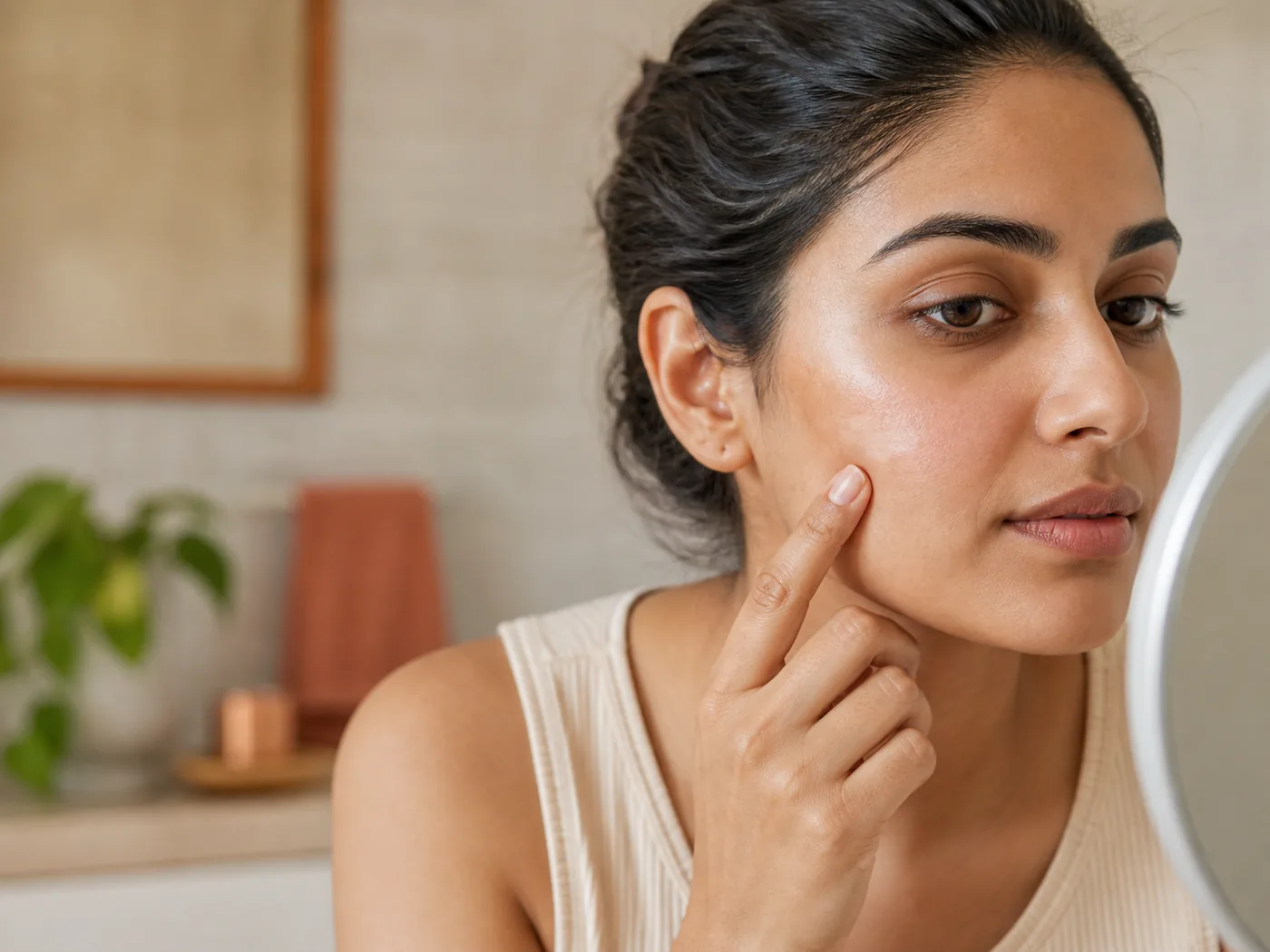
A flat pale patch, a tiny raised bump, and a flaky lighter area are different visual patterns. The goal is not to self-diagnose. It is to gather a clearer description so your next step is more sensible.
A flat patch can be related to pigment changes, dryness, irritation, or other causes. Track whether it is growing or changing.
A small firm bump may belong in the milia or clogged-bump family, but a photo alone should not be treated as a diagnosis.
Texture, itch, scale, or redness changes the urgency. A simple beauty routine may not be the right first move.
The best use of this page is to slow the decision down. Search intent around white spots on face often jumps straight from a visible concern to a product, procedure, or home remedy. A single image should not carry that much pressure. Use the sections below to name what you can actually see: color, border, texture, location, spread, symptoms, and timing. Then decide whether the next step is a calmer routine, a cleaner scan photo, a watch-and-track period, or a clinician’s opinion.
MyMirror is most useful when you treat it as a structured note, not a final answer. The scan can help you compare visible facial areas and avoid changing five products at once. It cannot diagnose, prescribe, confirm an infection, rule out a medical condition, or replace urgent care. If the concern is painful, rapidly changing, spreading, bleeding, infected-looking, near the eyes or lips, or affecting your daily life, use the photo record as context for a professional conversation rather than as permission to keep experimenting.
White spots on the face are especially easy to misread because “white” can mean several visual patterns: a flat lighter patch, a dry scaly area, a tiny raised bump, or a sharp depigmented spot. Some causes are harmless and temporary, some relate to eczema or dryness, some are pigment disorders, and some need treatment. The practical first step is not to diagnose from a selfie. It is to describe whether the area is flat or raised, smooth or scaly, stable or spreading, and whether itch, redness, pain, or sun sensitivity is present. That description makes your next step calmer and keeps you from scrubbing or bleaching a patch that may simply need gentle care or clinical evaluation.
| Signal | What it may look like | How MyMirror helps |
|---|---|---|
| Flat pale patch | A lighter patch may be hypopigmentation after irritation, eczema, sun contrast, or another pigment change. | The scan can help map location and compare whether the border is soft, sharp, growing, or stable. |
| Dry or scaly patch | Dryness, eczema-related changes, and pityriasis-alba-like patches can look lighter, especially after surrounding skin tans. | Texture notes help separate color-only concerns from barrier or irritation concerns. |
| Tiny raised bump | A pearly raised white bump may be more like milia or a clogged bump than a pigment patch. | Raised bumps should not be squeezed just because they look white in a photo. |
| Sharp white area | A very sharply defined white patch, especially if spreading, deserves professional assessment rather than routine guessing. | MyMirror can help you document progression, but diagnosis needs a clinician. |
Search results often collapse different skin patterns into one phrase. Separating lookalikes keeps the page useful and helps you avoid using a treatment meant for a different concern.
Often described as lighter, sometimes dry or scaly patches, commonly in children and teens. It is usually harmless but still worth discussing if new or spreading.
Usually sharper depigmented areas caused by pigment loss. A photo cannot confirm it, but clear spreading white patches should be assessed.
A yeast-related condition can create lighter, darker, pink, or brown patches and may include fine scale. It needs different care than cosmetic brightening.
Skin can look lighter after eczema, injury, acne, burns, or procedures. The timing after irritation is an important clue.
Raised dots are not the same as flat patches. The first decision is whether the white mark has height, firmness, or an opening.
For white spots, the safest routine avoids two common mistakes: scraping at the spot and trying to darken or bleach it. Gentle barrier care, sun protection, and pattern tracking are more useful while you decide whether a clinician should look at it. If there is scale, itch, spreading, or a sharply white border, treat it as a medical-clarity question instead of a product-shopping question.
Use a mild cleanser and a simple moisturizer. Dry or irritated skin can make lighter patches look more obvious, and strong exfoliation can make the area angrier.
Sun protection reduces contrast between surrounding skin and the lighter area. It also protects pale patches that may burn more easily.
If the mark is a small bump, squeezing can cause inflammation, scabbing, infection, or darker post-inflammatory marks.
Take a same-light photo every few weeks. If the patch is enlarging, multiplying, changing shape, or affecting confidence, bring that record to a clinician.
Different white-spot causes need different care. Using the wrong treatment can hide clues or irritate the face.
Use a scan to map where the lighter areas appear and whether they look flat, raised, or texture-led.
A scan cannot distinguish vitiligo, fungal changes, pityriasis alba, post-inflammatory color loss, or other conditions with certainty.
Avoid squeezing, scraping, or bleaching the area. Use gentle care, daily sun protection, and get medical advice for spreading or symptomatic patches.
MyMirror is designed to help with visible organization, not medical certainty. Use the photo to make a calmer next choice, then escalate when the skin itself asks for it.
Seek professional care if white patches spread, itch, hurt, scale heavily, appear after a new medicine, or affect confidence enough that you need a clear diagnosis.
No. Milia are raised tiny cyst-like bumps. Flat pale patches have a different set of possibilities.
No. MyMirror can organize visible patterns, but diagnosis belongs with a dermatologist.
Do not treat first and identify later. If the area is irritated, exfoliation can make it worse.
No. White or lighter spots can come from several skin patterns, including eczema-related changes, pityriasis alba, vitiligo, tinea versicolor, milia, or post-inflammatory color change. A vitamin deficiency should not be assumed from a face photo.
Yes, surrounding skin can tan while the lighter patch stays pale, making the contrast stronger. Some lighter patches may also be more sun-sensitive, so daily protection is a sensible starting point.
Usually no. Brightening products are designed for darker pigment, not unexplained pale areas. If the concern is actually pigment loss or irritation, brightening actives may make the skin more sensitive.
Many causes are not contagious, but some conditions need specific treatment. Because a photo cannot distinguish every cause, spreading or scaly patches are better checked by a clinician.
Any new, spreading, itchy, scaly, or confidence-affecting patch is worth asking a pediatrician or dermatologist about. The goal is reassurance and the right care, not alarm.
This page uses the provided Google keyword stats export for topic selection and cautious public dermatology references for safety boundaries: Cleveland Clinic on pityriasis alba and Cleveland Clinic on vitiligo and Cleveland Clinic on hypopigmentation.
Start with what is visible today, choose one sensible next step, and get professional care when the concern is painful, persistent, spreading, or worrying.
Start your free skin analysis now