
Post-Inflammatory Erythema (PIE) on Indian skin refers to lingering pink, red, or violaceous marks caused by damaged, dilated microscopic capillaries (blood vessels) near the skin surface following an inflamed pimple or squeezing trauma. Unlike brown post-inflammatory hyperpigmentation (PIH), which is driven by melanin, PIE is purely vascular. It blanches white under the pressure of a transparent glass tumbler. Applying exfoliating acids like glycolic acid damages fragile micro-vessels, triggering secondary hyperpigmentation. Dermatologists resolve PIE using vascular-soothing topicals (Centella Asiatica, 3%–5% Tranexamic Acid, 10% Azelaic Acid) paired with physical 100% Zinc Oxide mineral sunscreen.
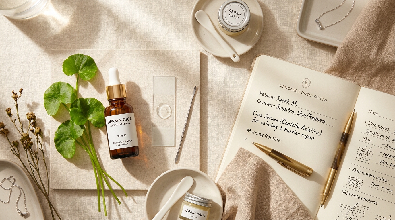
What Exactly Is Post-Inflammatory Erythema (PIE)?
When an acne bump appears on your cheek or jawline, an intense inflammatory battle occurs inside the pore. White blood cells flood the area to destroy trapped bacteria. To deliver these immune cells, your body opens up microscopic blood vessels called dermal capillaries.
If the pimple was deep, inflamed, or squeezed by fingers, the delicate walls of these tiny capillaries rupture and stretch. Even after the infection clears and the pimple flattens out, those tiny blood vessels remain stretched and engorged with oxygenated blood.
Think of a tiny rubber garden hose carrying fresh water beneath garden soil.
If someone steps hard on the hose, the rubber stretches out of shape and water pools in one spot, creating a puddle right beneath the surface. PIE is that exact pool of red blood trapped in a stretched-out micro-capillary. You are seeing red blood through translucent skin layers, not a stain of color on top of the skin.
Because Indian skin naturally produces abundant melanin, many people assume every post-pimple mark is brown pigmentation. But if your mark looks bright pink, cherry red, or purplish, you are dealing with vascular pooling, not melanin deposits.
The 5-Second "Glass Tumbler Test" (Diascopy)
Dermatologists use a simple clinical technique called diascopy to instantly tell whether a red mark is vascular or pigmentary. You can perform this test in your bathroom mirror in five seconds using any clean, transparent glass drinking tumbler or clear glass slide.
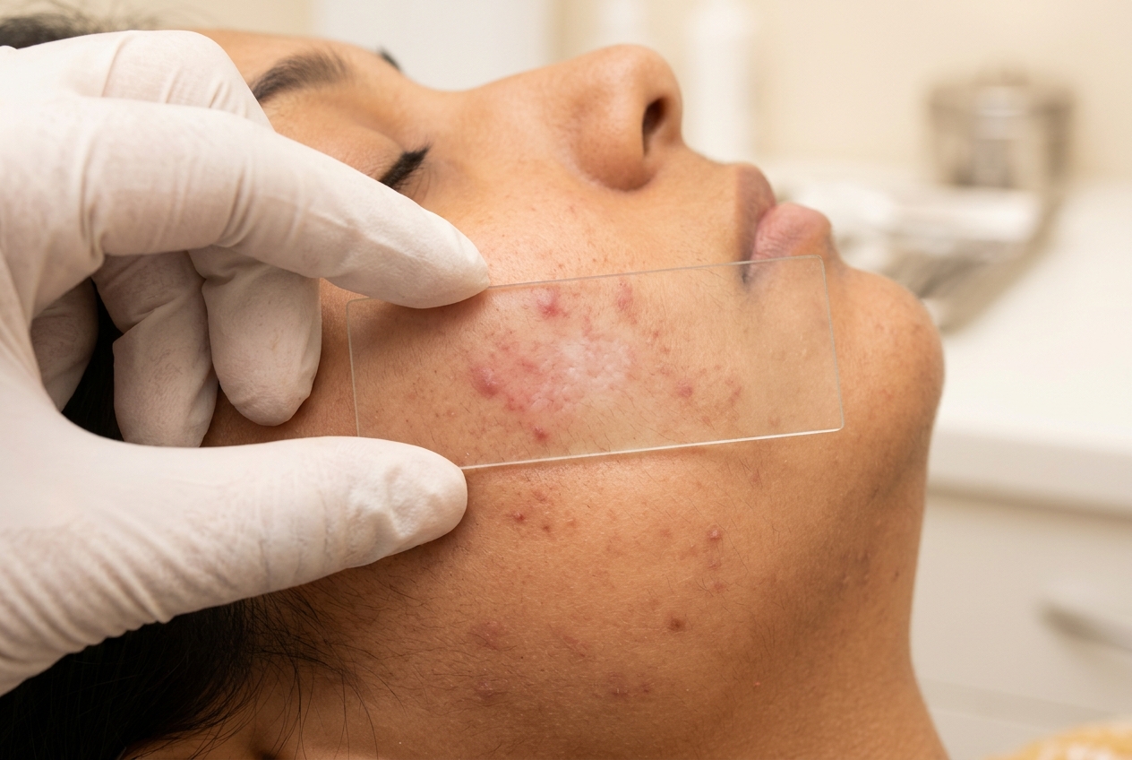
How to Perform the Test:
- Wash your hands and take an ordinary, clean transparent glass tumbler.
- Stand in front of a well-lit mirror and press the flat base or side of the glass firmly against the mark for three seconds.
- Look closely through the glass while keeping the pressure steady.
| What You See Through the Glass | Clinical Diagnosis | What Is Happening Under the Skin |
|---|---|---|
| The mark completely disappears or turns white (Blanches) | Pure PIE (Vascular) | The physical pressure temporarily pushed pooled red blood out of the dilated capillaries. |
| The mark stays brown, tan, or black (No change) | Pure PIH (Melanin) | The mark is made of solid melanin pigment granules deposited in skin cells, which cannot be moved by pressure. |
| The redness fades, but a faint brown ring remains | Mixed PIE + PIH | Both damaged blood vessels and stimulated pigment cells are present at the same time. |
Why Exfoliating Acids Disastrously Backfire on Red Acne Marks
When people notice an acne mark, their first reaction is often to grab an exfoliating acid: 10% Glycolic Acid toner, Salicylic Acid peel, or a high-strength AHA peeling solution.
On brown melanin spots (PIH), gentle exfoliation can assist cell turnover. But applying strong exfoliating acids to fresh, vascular red marks (PIE) causes serious damage to Indian skin.
If your red mark still blanches under glass pressure, stop all harsh peeling solutions, scrubs, and alcohol-based toners immediately. Your skin does not need exfoliation; it needs vascular repair and barrier soothing.
PIE vs PIH: The Complete Comparison for Indian Skin
Use this side-by-side clinical reference to understand how vascular redness and melanin hyperpigmentation differ in cause, behavior, and proper treatment:
| Characteristic | Post-Inflammatory Erythema (PIE) | Post-Inflammatory Hyperpigmentation (PIH) |
|---|---|---|
| Underlying Cause | Damaged, dilated microscopic capillaries (blood vessels) pooling hemoglobin. | Overactive melanocyte cells producing excess melanin pigment. |
| Visual Appearance | Bright pink, cherry red, or light violaceous purple spots. | Tan, light brown, dark chocolate brown, or slate gray spots. |
| Glass Test (Diascopy) | Blanches white under direct glass pressure. | Does not blanch; stays brown or dark under pressure. |
| Common Triggers | Squeezing or popping pimples, deep cystic swelling, skin picking. | Sun exposure on acne lesions, friction, post-peeling irritation. |
| Natural Healing Time | 8 to 24 weeks without treatment. | 6 to 18 months without treatment on Indian skin. |
| Hero Topical Actives | Centella Asiatica (Cica), Tranexamic Acid, Azelaic Acid, Niacinamide. | Kojic Acid, Alpha Arbutin, Glycolic Acid, Retinoids. |
| Fatal Mistake | Applying harsh chemical peels or scrubs that irritate blood vessels. | Skipping daily sunscreen, allowing UV rays to darken pigment. |
The 4 Proven Actives That Heal Microvascular Redness
To clear red acne marks, your routine must focus on two goals: calming inflamed blood vessels and reinforcing the capillary walls so they can shrink back to normal size. These four ingredients deliver the highest clinical success:
1. Centella Asiatica (Cica / Madecassoside) — The Capillary Healer
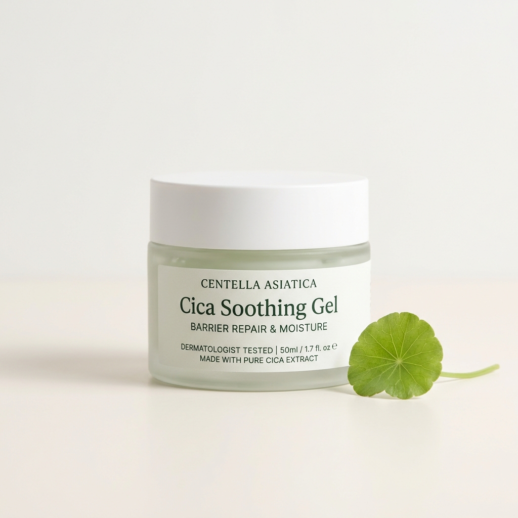
Centella Asiatica is a time-tested botanical extract containing four active triterpenoids: Madecassoside, Asiaticoside, Madecassic Acid, and Asiatic Acid. These compounds stimulate type I collagen synthesis around fragile microvascular walls, allowing stretched capillaries to regain elasticity. It provides immediate soothing relief without any risk of skin peeling or redness flares.
2. Tranexamic Acid (3% to 5%) — The Vascular Calmer
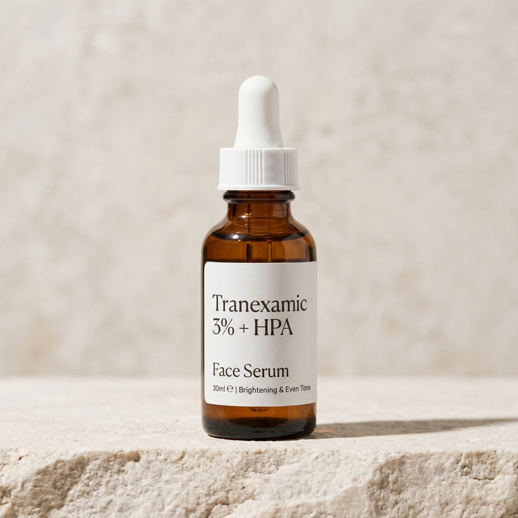
Originally utilized in medical settings to manage bleeding, dermatologists discovered that topical Tranexamic Acid directly blocks the plasmin pathway. By suppressing plasmin-induced vascular dilation and reducing vascular endothelial growth factor (VEGF), Tranexamic Acid shrinks swollen micro-vessels. As an added benefit, it stops pigment cells from turning that red spot into brown melanin.
3. Azelaic Acid (10% to 15%) — The Dual-Action Champion
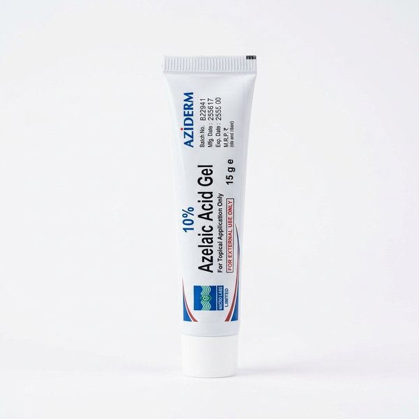
Available in Indian pharmacies as Aziderm gel or cream, Azelaic Acid is one of the most versatile ingredients for Indian skin. It reduces inflammatory swelling inside the microvascular network while simultaneously inhibiting tyrosinase, the enzyme that produces dark pigment. It is far gentler than glycolic acid and can be used on sensitive, inflamed skin.
4. 100% Zinc Oxide Mineral Sunscreen — The Thermal Shield
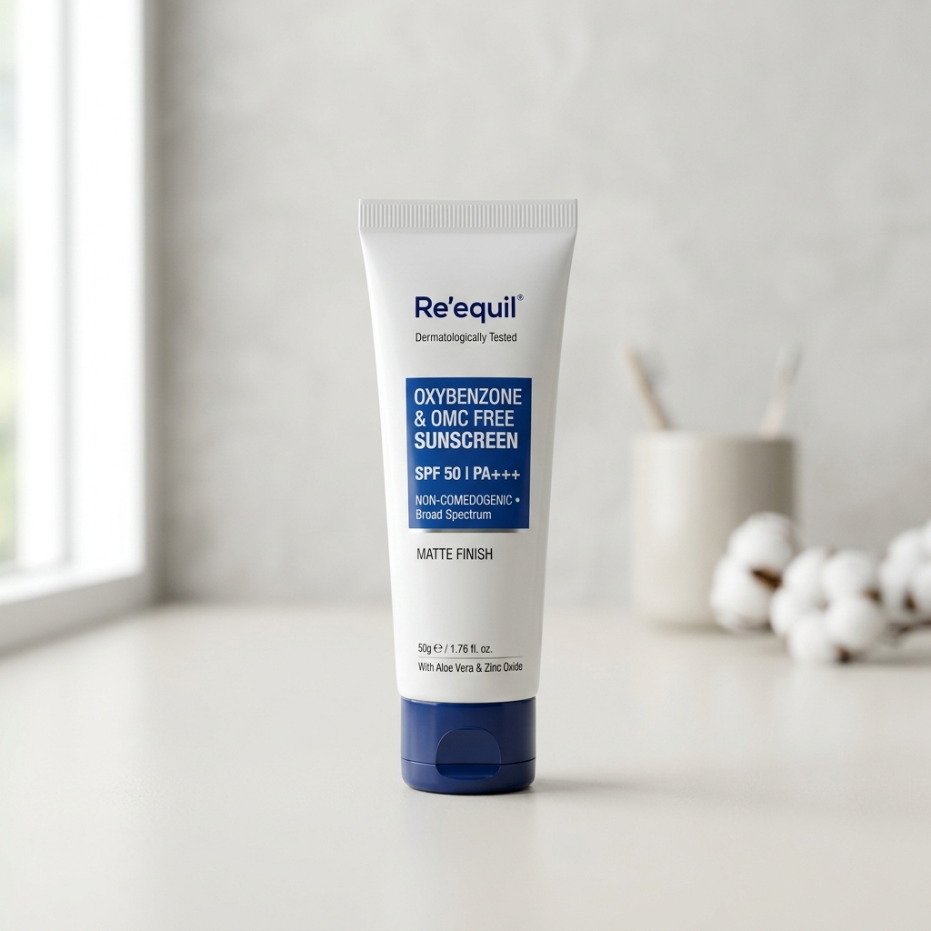
Chemical sunscreens absorb ultraviolet light and convert it into heat inside your skin. In Indian climates, this heat triggers facial vasodilation, making red spots flush brighter. A pure Zinc Oxide mineral sunscreen sits on top of your skin like a physical shield, bouncing away UV rays without generating heat. Zinc also acts as a natural anti-inflammatory agent that speeds skin recovery.
Dermatologist Video Guide: Treating Post-Acne Marks & Redness
Watch board-certified dermatologist Dr. Tanvi Vaidya break down why post-acne marks develop, how redness differs from pigmentation, and how to choose the right skincare strategy:
Clinical analysis by Dr. Tanvi Vaidya exploring post-acne vascular redness and hyperpigmentation management.
Your Week-by-Week Protocol for Fading Red Marks
Healing broken capillaries requires patience and consistency. Here is a realistic timeline of what to expect when you follow a gentle vascular-repair routine:
| Phase | Focus & Daily Routine | Visible Changes on Your Skin |
|---|---|---|
| Weeks 1 – 2 (Cooling & Calming) | Gentle non-foaming cleanser + Cica soothing gel + Zinc Oxide mineral sunscreen every morning. Zero scrubs or exfoliating acids. | Throbbing heat subsides. Bright angry red spots soften to a muted pink. Surrounding skin barrier strengthens. |
| Weeks 3 – 6 (Vascular Repair) | Introduce 3% Tranexamic Acid serum or 10% Azelaic Acid on alternate nights over moisturizer. Continue strict daily mineral sun protection. | Capillary pooling shrinks. Red spots lighten visibly in natural daylight. Positive diascopy blanching becomes faint. |
| Weeks 7 – 12 (Full Resolution) | Consistent use of Tranexamic Acid and Azelaic Acid. Spot-treatment on lingering marks. | Most superficial red spots fade completely into natural skin tone. Zero transition into dark brown PIH stains. |
Pharmacy Essentials & Calming Serums for Red Acne Marks
Here are four dermatologist-approved, non-irritating products available in Indian pharmacies and verified stores to heal vascular redness safely:
Frequently Asked Questions About Post-Acne Red Marks (PIE)
PIE (Post-Inflammatory Erythema) is vascular redness caused by damaged or swollen microscopic blood vessels near the surface of your skin after a pimple heals. It appears pink, red, or light purple. PIH (Post-Inflammatory Hyperpigmentation) is caused by excess melanin pigment produced by melanocytes, appearing tan, brown, or black. You treat PIE with vascular-soothing ingredients like Cica and Tranexamic Acid, whereas PIH requires melanin-inhibiting ingredients.
The glass test (clinically known as diascopy) uses an ordinary clean transparent glass tumbler or slide. Press the flat side of the glass firmly against the mark for 3 seconds while looking in a mirror. If the spot temporarily turns white or disappears completely while pressed, it is PIE because the pressure pushes pooled blood out of the dilated capillaries. If the mark stays dark or brown under pressure, it is PIH because melanin pigment is fixed in your skin cells.
Exfoliating acids like 10% Glycolic Acid or 30% AHA peels are designed to shed outer dead skin cells. But a red acne mark is an internal microvascular injury, not dead skin buildup. Applying strong exfoliating acids burns the delicate endothelial cells lining your healing capillaries, causing intense irritation. In Indian skin, this chemical trauma alerts melanocytes to deposit excess pigment, turning an easy-to-heal red vascular mark into a stubborn dark brown stain that takes months to fade.
Without treatment, mild red marks usually take 8 to 24 weeks to resolve as the body naturally repairs damaged capillaries. With a targeted routine containing Centella Asiatica, Tranexamic Acid, and daily mineral sunscreen, most individuals notice significant fading within 4 to 8 weeks. Severe or squeezed lesions that created deep vascular pooling may take longer or benefit from clinical vascular laser sessions.
A 100% Zinc Oxide mineral sunscreen is substantially better for red acne marks. Chemical sunscreens absorb ultraviolet light and convert it into heat energy inside your skin, which can cause micro-capillaries to dilate further and worsen facial flushing. Zinc Oxide acts like a physical mirror on your skin, bouncing UV rays away without generating exothermic heat and calming underlying inflammation.
Peer-Reviewed References
- Bae-Harboe YS, Graber EM. Easy as PIE (Postinflammatory Erythema). J Clin Aesthet Dermatol. 2013;6(9):46-49. PubMed: 24062874
- Mathews-Roth MM, et al. Postinflammatory erythema: clinical overview and management. J Cutan Aesthet Surg. 2020;13(1):1–7. PubMed: 32372791
- Lindgren AL, et al. Tranexamic acid in dermatology: a review. Int J Dermatol. 2021;60(11):1320-1327. PubMed: 33742440
- Park KY, et al. Vascular characteristics of post-acne erythema and therapeutic response to vascular lasers. Dermatol Surg. 2016;42(8):962-968. PubMed: 27391444
Unsure If Your Spots Are Red (PIE) or Brown (PIH)?
Take our free 60-second AI skin diagnostic scan. Our clinical tool analyzes your facial redness depth, detects underlying vascular inflammation, and gives you a customized dermatologist-reviewed routine before you spend money on wrong products.
Scan My Skin Free Now →